
How small can we go?
Beyond light: looking with electrons
This text has been automatically translated from Dutch with Google Translate.
Last year, Utrecht University bought a new device the size of two stacked cars. Why? To be able to look even better at particles of 100 nanometers (10,000 times as thin as a hair) in size. The new device is currently the best electron microscope in the world. One year after construction started, the new microscope is finally ready and can be used immediately for pioneering research in various fields. But how does such a microscope actually work? And why are those small particles so interesting?
In my search for answers to these questions, I speak with Savannah, a PhD student at Utrecht University and one of the few people who is allowed to use the new microscope for her research into the effect of Nickel catalysts. Savannah has been working with electron microscopes for about 8 years and knows her way around the field: “When people want to look at something small, they come to me”.
Let there be light!
Before we can understand an electron microscope, we first need to know how a normal microscope works. The very first microscope dates from the 17th century and is in the name of the Delft scientist Antoni van Leeuwenhoek. The device is basically a magnifying glass with an adjustable holder for viewing specimens.
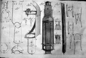
Figure 1: Schematic drawing of the first microscope developed by Antoni van Leeuwenhoek. Source: Wired
We now know the device as the first light microscope. That means that the microscope uses visible light to magnify with a lens. This makes your specimen appear larger when you look through the lens. The magnification of the lens depends on the degree to which the lens refracts the light. The more the light is refracted, the stronger the magnification.
Breaking light, won't it break?
Light can best be seen as a bundle of light waves (photons) that move in a certain direction. When the light beam collides with other material, this direction is adjusted. The thickness of the material is of great importance here. The light beam is slightly deflected at the transition from air to glass. Lenses make smart use of this by having different thicknesses along the length. As a result, the light is deflected more in the thick part than in the thin one
Modern microscopes still work exactly the same as those of Van Leeuwenhoek, but nowadays microscopes contain much better lenses to get even greater magnifications. Or, if we turn it around, to be able to look at smaller particles!
Light microscopes have been used extensively to study small things since their invention. In biology, microscopes are widely used to look at cells, almost everyone has used a light microscope in biology class. Everyone has only the greatest application of microscopy in their pocket: your cell phone! Not your phone itself, but the chips in the phone are so small these days that manufacturers have to use microscopes to properly check and mount the chips in your phone.
However, science is not sitting still and we soon ran up against the limit of light microscopy. The limit is not determined by the microscope itself, but rather by the light waves that need to be magnified. The light waves have a certain wavelength, with visible light it is between 400 nm (blue) and 700 nm (red). However, if we have a particle that is smaller than half that wavelength, the light waves will interact, which will lead to one blurry spot. So a bacterium can just be seen with a light microscope, but a virus is too small. So we had to look for something with a smaller wavelength.
Small smaller smallest
On campus, Savannah takes me to the microscopy department. We walk into a room where I immediately see a gigantic device. The microscope looks like a big white cupboard, but when a door opens, a small factory appears. The room is also crammed with servers, cables and other equipment. The new microscope barely reaches the ceiling and we can barely walk past it. There was no room for a computer anymore, so it has to be controlled from the outside. Savannah tells:
“Electron microscopy is very important for companies. Electron microscopy is used a lot in research into catalysts, (building) materials, medicines and even with the Corona vaccine. Companies have to rely on universities, because the cheapest is still as expensive as a new Maserati. Fortunately, they are getting cheaper and more user-friendly, so I expect it to be used more and more in the industry!”

Image 2: On March 1, 2021, the new Spectra300 microscope was put into use by Utrecht University. An official certificate proves that Utrecht University now has the best electron microscope in the world. Source: UU.nl
To increase the resolution of our microscope, a “light source” with a shorter wavelength is needed. Electrons are extremely suitable for this, because they have a wavelength of the order of picometers (0,000,000,000,001 meters). That is enough to even image atoms. Savannah tells:
“Because the wavelength of an electron depends on its energy, we can generate increasingly smaller wavelengths. The energy required for this can become so high that your sample breaks. Funnily enough, that is also an interesting property for research into new materials.”
As with light microscopes, we are already reaching the limits of electron microscopy. Savannah explains:
“The disadvantage is that we have to view the samples under ultra-high vacuum. In combination with the high energy of the electrons, this results in different properties of our samples compared to the properties at room temperature and air pressure. I expect that in the future these obstacles will also be overcome and that we can add gases or liquids to the samples. We can then look even more closely at our materials under the same conditions and in known reactions, which will make electron microscopy even more important.”
Utrecht University expects to move all microscopes to a new building by the end of 2021. A room has already been set up there for the next microscope, which is expected to be one car higher.