
21st Century Doctors
Human organs out of a 3D printer
My little brother was born with a heart defect. Fortunately, the doctors quickly found out what the problem was and surgery was enough to safe him. Unfortunately, surgery alone is not always enough. There are people who even need a whole new heart. You probably know someone as well who could even use a whole heart or perhaps a new pair of lungs. Now imagine that we can give those people a new organ. Doesn’t that sound great? Many researchers are also enthusiastic about this and made it their job. In the laboratory they 3D print small pieces of tissues that gradually resemble real human organs. Curious how they do this? Keep on reading!
You might be wondering now: why is it necessary to make human organs ourselves? Don’t we already have organ donors? You’re right. But the organ donors shortage is a major problem. For example, there are currently 127 people in the Netherlands on the waiting list for a new heart. This while there are only about 30 hearts available per year through organ donors. In order to get more organ donors, our government changed the Dutch donor law last July. This means that from your 18th birthday, “no objection to organ donation” will be registered behind your name. This of course unless you indicate you do not want this. Fortunately, this law change has reduced the shortage of organ donors. However, it is still a problem. A problem for which a whole new solution is on its way: 3D printing of organs.
How do these 3D printers work? Let’s take an example: what if we would like to print a heart valve. The printer first prints a thin layer of the bottom of the heart valve. This layer is printed with the ink that the researchers also call the “bioink”. The shape and size of the layer is determined by the g-code. The g-code is a setting that determines where exactly the bioink ends up in the 3D environment. Once this layer is finished, a new layer is printed on top of it. This continues until the top layer of the heart valve has also been printed.
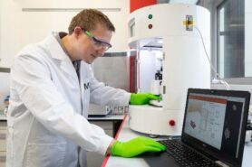
Researcher Jeroen Rouwkema experimenting with his 3D printer (Photographer: Rikkert Harink)
Who are the researchers who 3D print small pieces of tissue? These researchers work across different laboratories around the world. This includes the Dutch Jeroen Rouwkema, researcher and associate professor at the University of Twente. Last week I spoke to him and he told me more about his research. Jeroen Rouwkema: “My research group is involved in adding blood vessels to cultured tissues. These tissues have two different applications: the tissues are an alternative to donor tissue, or they are used as a test system for new drugs.”
In Jeroen Rouwkema’s research it is important to understand that blood vessels always adapt to their environment. This also happens with the organs of your body. For example, if you exercise more, your muscles will grow. These muscles then need more oxygen and nutrients. Your body responds to this by expanding the network of blood vessels in the muscle tissue. Jeroen Rouwkema uses this in his research as well: “We want to design the environment of the tissue in such a way that we can control the organization of the blood vessels.”
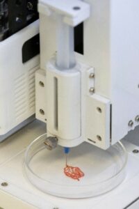
3D printed blood vessel network (Photographer: Rikkert Harink)
Jeroen Rouwkema experiences many advantages by using his 3D printer. The biggest advantage is that he can determine very specifically, in 3D(imensions), where the bioink is applied. This is not possible with other technologies. Jeroen Rouwkema says that it is also possible to add a fourth dimension: “The blueprint for the tissue is drawn in three dimensions and in the fourth dimension, which is time, you can determine what the tissue will look like”.
A 3D printed organ versus one from an organ donor has several advantages. For example, your own heart cells can be used when printing a new heart. In the case of a heart from an organ donor, you are dealing with other cells. And those cells can be very different from your own cells. This increases the chance that your body will not accept the donor heart. Another advantage is that the 3D printed tissues can also be used in research. For example, by testing new drugs. This reduces the need for animal testing.
Although the 3D printing of tissues and organs is still very much in development and important steps are being taken, there are still some challenges for the researchers. For example, many organs consist of many different cells. It is therefore difficult to properly combine all these different cells. Many organs also have a complicated structure. Did you know for example that one lung consists of 300 million small air bubbles? That will be difficult to print!
In addition to these challenges, researchers also sometimes have doubts about the use of the 3D printing technique itself. Jeroen Rouwkema: “There is a great hype factor in the research about the 3D printing technique. This technique is often used when it is not necessarily necessary.” He also said: “In reality, it may be very expensive to have a new heart printed. Who will pay for this? Is might only be available for the people who own a lot of money.”
Despite the challenges and doubts that remain, it is very exciting what this research field will look like in 100 years. Can we really print a new heart or lungs in 100 years’ time? Or have we already found another solution for the organ donor shortage by then? This is what Jeroen Rouwkema said about it: “I see the body as a collection of machines, each with a very specific function. For example, a heart is a pump that pumps blood around the body. Making something else that also works well as a pump is more important than something that resembles a natural heart as much as possible. “
Written by: Franca van Heijningen, Master Student Biomedical Sciences